 Italiano
 English
Detecting an object without regaining normal vision
A retina can lose the cells that collect light while retaining those that carry the message to the brain. An international team is trying to give the remaining cells a new task: becoming sensitive to light. In results announced on October 7, 2026, some participants with advanced retinitis pigmentosa regained limited abilities to detect or locate objects. This is the central idea: creating an alternative input for visual information when the natural one is severely damaged.
The combination includes gene therapy and special goggles. It does not yet allow people to read or recognize faces, but points to a different possibility from trying to preserve dying photoreceptors. For someone who has lost almost all vision, distinguishing where something is located is a practical question. We do not yet know how far this result can translate into independence beyond testing: that is precisely the step that will make subsequent research decisive.
The problem: the sensors stop working
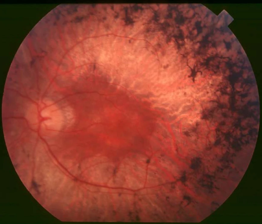
Retinitis pigmentosa encompasses genetic diseases that progressively damage the retina, the tissue at the back of the eye. Difficulties often begin in the dark and in peripheral vision; central vision can also diminish over time. The speed varies from person to person. Different genes can cause the condition, so describing a single disease and a single solution risks hiding important differences.
Photoreceptors, called cones and rods, convert light into signals. Retinal ganglion cells are part of the pathway that relays them to the brain. The idea behind optogenetics is to give light sensitivity to cells that normally perform another role. This is not the same as rebuilding the original retina: it modifies the route through which information can arrive. That distinction explains both the innovation and why the resulting vision remains very different from normal sight.
How it works, from the injection to the goggles
The first step is an injection into the eye. In the work published in 2021, the viral vector carried instructions to produce ChrimsonR, a light-sensitive protein. The instructions enabled ganglion cells reached by the treatment to respond to a light stimulus. The vector is the delivery vehicle: it does not replace lost cells and is not an electronic implant inside the eye.
The second step takes place in front of the face. A camera in the goggles detects changes in the brightness of the scene; the device turns them into light pulses projected onto the retina. Simply wearing more powerful ordinary glasses is therefore not enough. The system must translate what it captures into a signal that activates the treated cells in real time. In the first clinical case, the patient could perceive, locate and count objects using the treated eye and the device.
The third step concerns interpretation. The new signal must become useful to a person, rather than merely measurable in a laboratory. In the updated cohort, those who had practised more with the goggles tended to achieve better results in object detection. This is an observed association, not proof that a particular number of hours guarantees an outcome. It nevertheless explains why treatment needs to be considered together with rehabilitation, rather than as a single injection that solves everything.
What the ten-person study demonstrates
The team led by José-Alain Sahel of the University of Pittsburgh and Botond Roska of the Institute of Molecular and Clinical Ophthalmology Basel extended the earlier individual result to ten participants. According to the researchers’ account, six reached the predefined threshold for a clinically meaningful improvement in light sensitivity. Some improved at detecting or locating objects with the goggles. These are limited abilities, not evidence of fully restored sight.
Safety was the study’s main objective. The scientific press release reports mainly mild or moderate eye-related events and one severe event immediately after injection that resolved within minutes following treatment. Saying that no obstacles emerged that would stop this experimental path is different from declaring it risk-free. Ten people are not enough to characterize rare events or predict the outcome for all potential candidates.
The important test is what a person can do
A paper from April 2026 on methods for evaluating optogenetic therapies clarifies a problem often invisible in news coverage: recording electrical activity does not automatically mean useful perception. A response in the retina, a signal in the brain’s visual areas and recognizing an object are related measurements, but they are not interchangeable. Understanding the benefit requires several levels of observation, including behavior.
This distinction changes how we read the story. A biological signal demonstrates that something is activated; a successful task shows a possible use of that signal. The next step will be to understand how repeatable that use is, under which conditions and with what effort. These questions also concern the device: weight, simplicity and adaptation to everyday situations would be components of the value of a potential treatment, rather than details to address after the discovery.
Vision rehabilitation already exists to help people with sight loss carry out everyday activities. The National Eye Institute describes it as a combination of services, aids and learning strategies. The new technology does not erase that experience: it can add a different type of information to integrate. The interesting future therefore also includes professionals able to teach people to use it and trials designed around people’s needs.
What changes, and what still needs to be built
The shift is in the target: when natural sensors can no longer be used effectively, researchers seek a function in surviving cells. These results do not tell us whether the approach will become an accessible treatment in Italy, or what its overall cost would be. There is no verified price to attach to a future offering that would include therapy, a device, monitoring and training. Presenting it as an available cure today would be incorrect.
For Frontiera, the consequence to follow is the transition from laboratory performance to an ability chosen by the person who will use it. Success cannot be measured solely by the sophistication of the protein or the goggles. It must include the benefit relative to the effort required, its duration and the people for whom it works. Change takes shape when a technology meets a real need; here, that need is to use visual information that previously did not arrive.